|
Clinical
Services

-
Consulting
-
Paraclinic Evalution
-
Topography
-
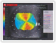
Orbscan


VuMax II /Sonomed.Inc.
High Resolution Ultrasound
Biomicroscope :
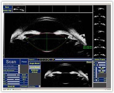
The VuMax-II, Ultrasonic
Bio-Microscope takes you to
another level of accuracy in
high resolution, high
frequency ultrasound with
its unmatched image
resolution and precise
sulcus to sulcus and high
detail angle imaging. Users
can now visualize structures
and mechanisms behind the
Iris that cannot be seen
with OCT technology.
The VuMax-II also
incorporates powerful
processing tools and our
proprietary image
enhancing Focus Software
enabling users clearer,
sharper details of images
previouslynot attainable.The
systems is PC-based,
networkable and is capable
of AVI and JPEG file formats
for export.
The 18.5mm x 14 mm deep
scanning field captures the
entire anterior
segment in one scan and
provides advanced
intraocular measurement
capabilities.
The 45-second digital
dynamic recording capability
allows playback, adjustment
of
Gain,TGC and Contrast, live
zoom scan and isolation of a
single frame for more
efficient
examination and better
diagnosis.
The VuMax-II comes with a
lightweight, hand-held probe
and three custom made
immersion cups to ensure a
comfortable fit and ease of
use. Non-invasive
evaluations can
be preformed anywhere that
the patient can be
comfortably reclined.kly and
easily on an Ultrasound
biomicroscopy utilizes high
frequency transducers to
obtain high resolution. Our
unit, with a 35 MHz
transducer, achieves a
resolution of approximately
50 microns, and has a tissue
penetration of 5.0 mm. In
vivo, cross-sectional or
transverse images can then
be obtained
detailing the cornea, iris,
ciliary body, anterior
chamber angle, and
peripheral sclera
to demonstrate structural
relationships.
Ultrasound Biomicroscope
Applications
-
Glaucoma Management
-
IOL and Phakic IOL Lens
Implantation
-
Accomodative IOL
-
High Resolution Imaging
of Anterior Segment
-
B-Scan Imaging of
Posterior Segment
Normal Eye Normal Eye
UBM Image
Angle-Closure Glaucoma
Pupillary block
Before and after laser
iridotomy
Dark room provocative
testing
Plateau iris
Iris cysts and tumors
Pigment Dispersion Syndrome
and Pigmentary Glaucoma
Before and after laser
iridotomy
Before and during
accommodation Concave Iris
Configuration in Pigment
Dispersion Syndrome

Ocular Trauma
Cyclodialysis cleft ,Angle
recession
Intraocular Lens Position
Capsular bag fixation
Sulcus fixation
Malpositioned IOL
Failing Filtering Blebs
Functioning blebs
Four sites of obstruction to
outflow
The VuMax Ultrasound
Biomicroscope can display
and capture the entire
anterior segment in motion,
helping surgeons to
visualize and better
understand lens placement
and dynamic movement. These
motion clips can be reviewed
frame by frame in high
detail, providing an
invaluable analysis tool.
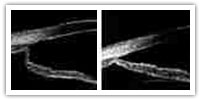
Accommodation and Iris
Configuration

An increase in the iris
concavity can be induced
during accommodation (right)
when compared to the
non-accommodative state
(left).

LS 900 for IOL Calculation

|